Search This Blog

Sunday, November 21, 2010

Oxidation of Dopamine


Parkinson's disease is characterized by the progressive neurodegeneration of neuromelanin containing neurons in the substantia nigra pars compacta of our brains. One of the mechanisms proposed for the selective degeneration of dopaminergic cells is the natural tendency of dopamine to become oxidized and induce a chain reaction of oxidative stress inside the cells. The reactive oxygen species as well as the end products of this reaction facilitate the formation of Lewy bodies when accompanied with an intraneuronal accumulation of ubiquinilated alpha synucleins.


The oxidation of dopamine produces dopamine o-quinone, a melanin precursor. This reaction is mediated by Tyrosinase, an enzyme that catalyzes the oxidation of o-diphenols, such as dopamine, into o-quinoles using dioxygen. In pathological conditions, a reactive oxygen species is generated, namely dopamine o-semiquinone, and its formation is catalyzed by lactoperoxidase and hydrogen peroxide. Pathological conditions may involve the enzymatic breakdown of monoamine oxidases (MAOs) and/or the induction of oxidation reactions from environmental toxic agents. Monoamine oxidases normally recycle extra amounts of dopamine in the brain. The end product of the oxidation of dopamine is an Aminochrome that carries a detrimental function in parkinson's disease as it has been shown to damage essential cellular macromolecules and increase the production of ROS.


Briefly, Aminochrome undergoes a one-electron reduction reaction where redox cycling DA-semiquinones are formed. The enzyme that mediates the reduction of Aminochrome to the o-semiquinone is the NAPDH cytochrome P450 reductase. Dioxygen species are oxidized to ROS while NADPH to NADP. Interestingly, antioxidants such as Superoxide Dismutase and catalase exacerbate this reaction since it depletes the cell fr
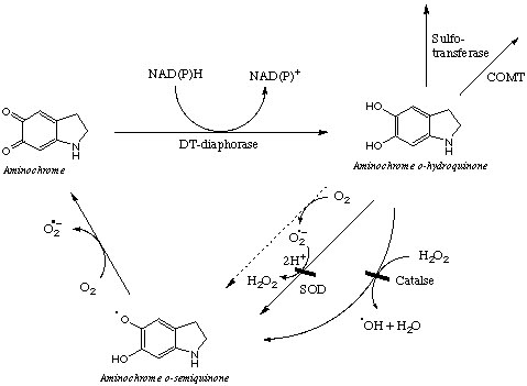
om superoxide species, thus moving the reaction towards the reactant (aminochrome). Thus, the presence of SOD and catalase increases the auto-oxidation of Aminochrome o-semiquinone. However, in the presence of DT-diaphorase, as it is shown in the right, Aminochrome is reduced to Aminochrome o-hydroquinone. Although o-hydroquinones are also auto-oxidized; the rate of auto-oxidation is lower than that of aminochrome o-semiquinone. One of the possibilities is that SOD and Catalase recognize a general motif in the structure of the o-hydroquinone and prevents the generation of the degenerative o-semiquinones. Interestingly, the alcohols are present in the benzene rings of dopamine and o-hydroquinone. The cyclilization of the amine group is the only feature that differs among dopamine and aminochrome o-hydroquinone. The hydroquinone is a stable structure that is depleted from cells by reactions mediated by sulfo-transferases and COMT.

Question: How pertinent is the expression of DT-diaphorase for the progression of parkinson's disease, do these patients have a lower expression of DT-diaphorase in the substantia nigra? Do dopaminergic cells that express high levels of DT-diaphorase in the brain less likely to die from the disease?

Fast-scan cyclic voltammetry for dopamine detection

Bloq question: How can we study changes in dopamine concentration inside cells in vivo?

The principle behind this technology is the readily oxidation of dopamine in air and the unique transfer of electrons to a microelectrode as dopamine changes into its oxidized form (o-quinone).

Fast-scan cyclic voltammetry (FSCV) is a modified method of the classic cyclic voltammetry technique that is used to detect electroactive molecules based in changes in their redox potentials. FSCV provides a much higher temporal resolution at a subsecond time scale for analyte detection. The carbon-fiber microelectrodes monitor the extracellular concentration of electroactive molecules and are suitable for the in situ and in vivo monitoring of dopamine concentration inside dopaminergic cells in awake animals or in single cells.

The potential at the microelectrode is held at a voltage below the oxidizing potential (-0.4 V in reference to a silver/silver chloride electrode) of dopamine and it is linearly ramped to an oxidizing potential (+1.3 V) and back. During this cycle, characteristic of a redox reaction, electrons are transferred between the microelectrode and dopamine-o-quinone at the positive sweep and back to dopamine in the negative sweep. The scan rate of the microelectrode is held at a high rate and it is set to measure the flux of electrons (as current) multiple times each second. Current is proportional to the number of dopamine o-quinone molecules generated by the electro-oxidation reaction normalized to a standard. Current is plotted against potential to yield a cyclic voltammogram (CV) graph that is used to identify the analyte, since each electroactive molecule behaves differently during a redox reaction. The peak in the cyclic voltammeter plot is representative of dopamine o-quinone (~+1.3 V) and the change in the peak height is proportional to change in concentration when normalized to a standard.

FSCV permits the study of changes in the dopamine concentration over time by plotting the peak oxidation potential at different time points or over a range of biological preparations.

Reviews
Probing brain chemistry Stamford JA and Justice JB Jr Analytical Chemistry 68, 359A-363A (1996)


Critical guidelines for validation of the selectivity of in-vivo chemical microsensors Phillips PEM and Wightman RM Trends in Analytical Chemistry 22, 509-514 (2003)


Detection technologies. Probing cellular chemistry in biological systems with microelectrodes Wightman RM Science 311, 1570-1574 (2006)

Thursday, September 9, 2010

Biogenics in Neuroscience

Scientists at the University of Utah are working on many projects that aim to connect the functional activities of the brain to electrical circuits to solve sensorimotor problems.
http://www.bioen.utah.edu/faculty/greger/

Wednesday, July 28, 2010

Comparative Mammalian Brains


I just found this wonderful website for the comparative anatomical study of the brains in different animal systems. Enjoy!
http://www.brainmuseum.org/

Friday, June 25, 2010

Neuroscience Protocols

Please use this to share protocols of interest to you. Provide a purpose to the article and, if possible, comment on the role of the major ingredient(s) in your protocol. Thank you!

Thursday, June 24, 2010

Neuroscience Books

Name your favorite neuroscience books and write short book reviews. The purpose of this session is to update your fellow scientists and generate interest in the field of neuroscience to newcomers.

Monday, June 14, 2010

Functional Brain Atlas


Allen Human Brain Atlas (www.brain-map.org) has launched a human brain atlas spatially mapped with microarray data in over 700 distinct anatomical locations.
The Allen Mouse Brain Atlas also carries a genome-wide image database of gene expression with In Situ Hybridization and Nissl images. You can select a structure to select genes with high levels of induction and search for a gene in specific anatomical region or in the whole brain.

Sunday, June 13, 2010

Introductory remarks

Welcome to the homepage of our new discussion blog led by a group of neuroscientists at the IMM from UT Health. This blog is designed in lieu of forming a strong team of neuroscientists at the IMM and connecting us with other well-established programs and scientists at UT Medical School and other Institutions. We will discuss scientific publications and share novel protocols in the neurobiology of disease. We welcome your input to our discussions as well as other information relevant to the study and appreciation of the nervous system in healthy and diseased states . Thank you!